Mass Cytometry
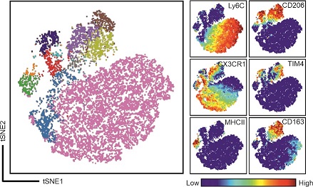
Like traditional fluorescence-based flow cytometry, mass cytometry allows multiple cellular parameters to be measured simultaneously. Instead of the in flow cytometry used fluorochromes, mass cytometry uses heavy metal reporter ions. This allows the quantification of up to 45 cellular parameters simultaneously with hardly any overlap. Thus, mass cytometry or cytometry by Time of Flight (CyTOF) adds new dimensions to the study of cellular heterogeneity and diversity.
Helios (CyTOF)

The Helios is a modern mass cytometer, which quantifies cell-associated heavy metals using inductively coupled plasma mass spectrometry. With an analytical range of 75-209 Dalton, it provides users with 135 detection channels that can simultaneously resolve multiple elemental probes at high acquisition rates superior mass resolution provide researchers with the ability to differentiate adjacent peaks.


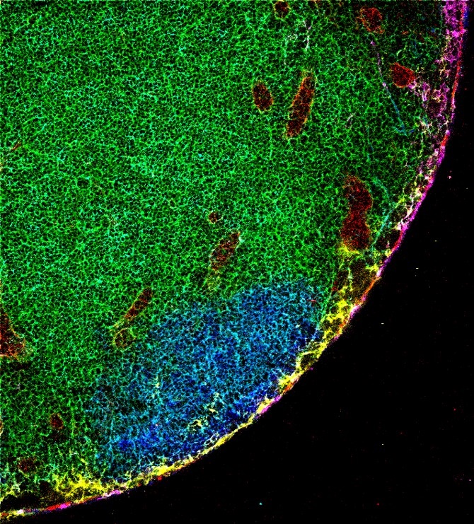
Imaging mass cytometry combines immunohistochemistry, using metal ions coupled antibodies and markers with mass cytometry (CyTOF) to rebuild an image of frozen or fixed tissue sections or stained cells on a microscopic slide.
Hyperion
This is the imaging unit, which combined with the mass cytometer Helios forms the platform for imaging mass cytometry.
The platform can simultaneously detect 4 to 37 protein markers in biological samples including fixed tissue sections or cells deposited onto glass slides (liquid biopsies). This allows researchers to investigate cellular subpopulations in various tissue microenvironments. The system allows for cellular profiling in spatial proximity, enabling subpopulation profiling and exploration of relationships of neighboring cells within the context of the tissue structure.
Simple workflow

Technical data
Technical data for our Mass Cytometry Instruments can be found on the webpage of Standard Biotools Inc. www.fluidigm.com. For Helios Here For Hyperion Here
Reviews on Mass Cytometry
Spitzer, MH. and Nolan GP. Mass Cytometry: Single Cells, Many Features; Cell 165, May 5, 2016 http://dx.doi.org/10.1016/j.cell.2016.04.019
Tanner, SD; Baranov VI, Ornatsky OI; Bandura DR; Geaorge TC. An introduction to mass cytometry: fundamentals and applications. Cancer Immunol Immunother (2013) 62:955–965 DOI: 10.1007/s00262-013-1416-8
Hartmann FJ and Bendall SC. Immune monitoring using mass cytometry and related high dimensional imaging approaches. Nature Reviews, Rheumatology Vol 16 Feb 2020. PMCID: PMC7232872; DOI: 10.1038/s41584-019-0338-z
Baharlou H, Canete NP, Cunningham AL, Harman AN and Patrick E: Mass Cytometry Imaging for the Study of Human Diseases—Applications and Data Analysis Strategies. Frontiers in Immunology Vol.10; Nov 2019. https://doi.org/10.3389/fimmu.2019.02657


